A Rare Case Report of Angina Bullosa Hemorrhagica
Dhanya Susan Eapen1, Ramiya Ramachandran Kaipuzha2, Davis Thomas Pulimoottil3, Nithin Mohan3
1. Junior Resident, Department of ENT, Al Azhar Medical College and Super Specialty Hospital, Thodupuzha, Kerala, India;
2. Registrar, Department of Otorhinolaryngology, Jahra Hospital, Al Jahra, Kuwait;
3. Department of ENT, Al Azhar Medical College and Super Specialty Hospital, Thodupuzha, Kerala, India*
Corresponding Author: Dr. Nithin Mohan,
Department of ENT, Al Azhar Medical College and Super Specialty Hospital, Ezhalloor P.O.,
Thodupuzha, Idukki District, Kerala - 685605
Phone: 9400635735, Email: nithinmohankedaram@gmail.com
ABSTRACT
Background: Angina Bullosa Hemorrhagica (ABH) is a rare, benign disorder characterized by the sudden appearance of blood-filled bullae in the oral mucosa without any identifiable systemic, hematologic, or autoimmune cause. We report a case involving a 66-year-old diabetic male who developed a solitary hemorrhagic blister on the right buccal mucosa shortly after eating. The lesion resolved spontaneously within 24 hours without medical intervention and showed no recurrence over a one-year follow-up. This case highlights the classical presentation of ABH and reinforces the importance of recognizing its self-limiting nature to avoid unnecessary investigations and treatments. A brief discussion of the clinical presentation, possible etiological mechanisms, differential diagnosis, and management is provided to increase clinician awareness of this often underdiagnosed condition.
Keywords: Angina Bullosa Hemorrhagica, Buccal mucosa, Blister, Chlorhexidine
Introduction
Angina Bullosa Hemorrhagica, first described by Badham in 1967,1 refers to the sudden appearance of subepithelial hemorrhagic bullae in the oral cavity in the absence of a systemic disease or blood dyscrasia. The condition derives its name from the classical angina-like suddenness and its hemorrhagic blistering appearance. ABH is clinically striking, often alarming both patients and physicians due to the appearance of large, blood-filled vesicles or bullae, yet it typically follows a benign and self-limiting course. These lesions most frequently affect the soft palate, buccal mucosa, and lateral aspects of the tongue. Despite its dramatic presentation, ABH is often underdiagnosed or mistaken for other serious vesiculobullous or hematologic conditions. Recognizing the typical presentation and benign course of ABH is essential to avoid extensive diagnostic workups and inappropriate treatment.
Case Report
A 66-year-old male, known to have well-controlled type 2 diabetes mellitus, presented with the sudden appearance of a dark swelling on the inner surface of his right cheek (figure 1). He noticed the lesion shortly after consuming solid food. There was no history of trauma, recent dental procedures, medication use such as anticoagulants or corticosteroids, or any systemic symptoms. He did not report pain, burning, bleeding from other sites, or difficulty swallowing.
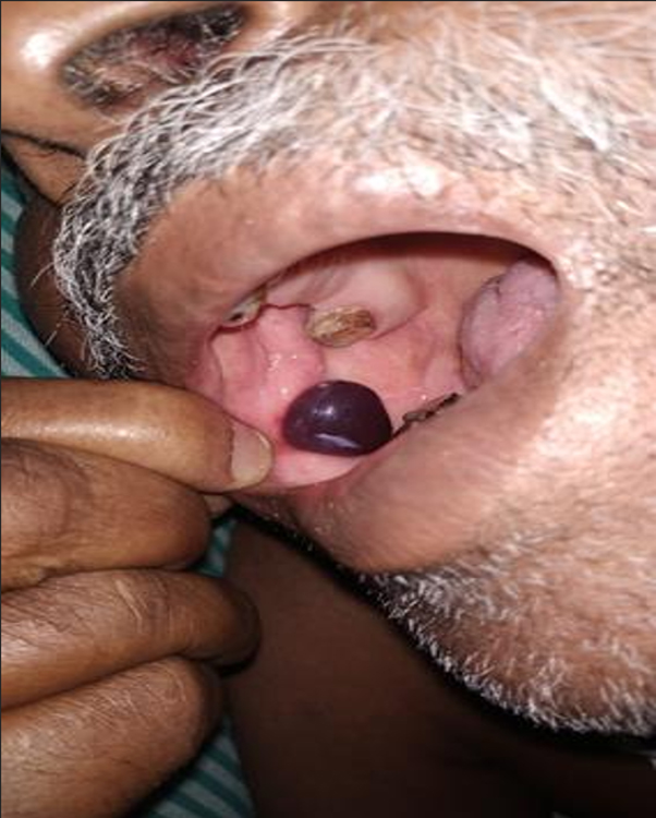
Figure 1. Clinical image showing a solitary, non-tender hemorrhagic bulla (3 × 2 cm) on the right buccal mucosa adjacent to the right mandibular second premolar.
On clinical examination, a tense, dark bluish-purple bulla approximately 3x2 cm in size was observed over the right buccal mucosa. The rest of the oral cavity was normal. There were no petechiae, purpura, or gingival bleeding. No lymphadenopathy or systemic findings were noted. A clinical diagnosis of Angina Bullosa Hemorrhagica was made based on the characteristic location, sudden onset, hemorrhagic content, and absence of systemic involvement. Given the typical presentation, no biopsy was performed.
The lesion ruptured spontaneously within a few hours, releasing blood and leaving behind a shallow erosion. The site healed over the next three days without any scarring (figure 2). The patient was managed conservatively with reassurance and advised to use chlorhexidine mouthwash for maintaining oral hygiene. He was not prescribed any specific medications. Laboratory investigations including complete blood count, platelet count, and coagulation profile were within normal limits. At a one-year follow-up, the patient remained asymptomatic with no recurrence of similar lesions.
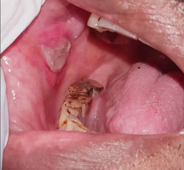
Figure 2. Post-rupture appearance of the lesion showing mucosal remnants and healing site.
Discussion
ABH is characterized by the rapid onset of blood-filled bullae on the oral mucosa in the absence of identifiable systemic disease or trauma significant enough to explain the lesion. The buccal mucosa, soft palate, and lateral tongue are the most commonly affected areas.2 These lesions usually appear spontaneously or following minor trauma, such as mastication, hot beverages, or minor dental manipulations. The lesions are typically painless, though some patients may report a sensation of fullness or mild discomfort. Affected blisters often rupture spontaneously within hours or days, and healing occurs rapidly without scarring. In the present case, the lesion healed within three days without any intervention.3
Although the exact pathogenesis of ABH remains unclear, minor trauma is considered the most likely trigger. In susceptible individuals, even trivial injury may cause subepithelial separation and hemorrhage.4 The role of systemic conditions like diabetes mellitus has been proposed, as microvascular changes and delayed wound healing may contribute to mucosal fragility. Inhaled corticosteroids are also considered potential risk factors due to their atrophic effect on mucosal tissues. Histopathologic examination, though seldom required, typically reveals a subepithelial cleft with blood accumulation and minimal or no inflammatory infiltrate, supporting a non-inflammatory etiology.5
Given its benign nature, ABH is largely a diagnosis of exclusion. However, the alarming presentation necessitates a thoughtful consideration of other potential causes. Vesiculobullous diseases such as pemphigus vulgaris, mucous membrane pemphigoid, and bullous pemphigoid should be ruled out. These autoimmune disorders often present with painful, persistent, and recurrent lesions involving multiple mucosal sites, and they typically require biopsy with direct immunofluorescence for definitive diagnosis. Hematological conditions such as thrombocytopenia, leukemia, or coagulopathies must also be considered, especially in patients with other bleeding manifestations or abnormal blood counts.6 Vascular lesions such as hemangiomas or localized angioedema may resemble ABH but tend to have a different clinical course and often lack spontaneous resolution.
Management of ABH is conservative. The primary approach is reassurance and observation, as the condition resolves spontaneously. Patients should be informed about the benign nature of the lesion to alleviate anxiety. Symptomatic management may include antiseptic mouthwashes such as chlorhexidine to prevent secondary infection and promote healing. Analgesics may be prescribed if there is discomfort, although pain is usually mild or absent. In cases where large bullae develop near the oropharynx or soft palate, there is a theoretical risk of airway obstruction. Such scenarios warrant careful monitoring and, in rare cases, incision and drainage under sterile conditions by an otolaryngologist to prevent airway compromise.
Preventing recurrence involves addressing contributing factors. Inhaler technique should be reviewed in patients using corticosteroids, and a spacer may be considered. Good glycemic control in diabetic individuals may also reduce mucosal fragility. Though vitamin C and bioflavonoids have been suggested as prophylactic agents, the evidence supporting their efficacy remains anecdotal and inconclusive.
The present case illustrates the classical features of ABH, including the sudden appearance of a solitary hemorrhagic blister, rapid resolution, and complete healing without intervention or recurrence. The absence of systemic involvement, normal hematological parameters, and the characteristic location of the lesion further supported the diagnosis. Timely recognition of ABH is crucial to prevent unnecessary investigations, biopsies, or empirical treatments.
Conclusion
Angina Bullosa Hemorrhagica is a rare but benign oral condition that should be considered in the differential diagnosis of sudden oral blood-filled blisters, particularly in elderly or diabetic patients. Awareness of its self-limiting nature and typical presentation can prevent misdiagnosis and avoid unnecessary treatment. A thorough history and clinical examination are often sufficient for diagnosis, and reassurance remains the cornerstone of management. This case emphasizes the importance of clinician familiarity with ABH to distinguish it from more serious conditions and to manage it effectively with a conservative approach.
END NOTE
Author information
- Dr. Dhanya Susan Eapen, Junior Resident,
Department of ENT, Al Azhar Medical College and Super Specialty Hospital, Thodupuzha, Kerala, India.
Email: dhanueapen@gmail.com - Dr. Ramiya Ramachandran Kaipuzha, Registrar, Department of Otorhinolaryngology, Jahra Hospital, Al Jahra, Kuwait
Email: ramiyaramk@gmail.com - Dr. Davis Thomas Pulimoottil, Professor, Department of ENT, Al Azhar Medical College and Super Specialty Hospital, Thodupuzha, Kerala, India.
E-mail: davisthomasp@yahoo.in - Dr. Nithin Mohan, Assistant Professor,
Department of ENT, Al Azhar Medical College and Super Specialty Hospital, Thodupuzha, Kerala, India.
E-mail: nithinmohankedaram@gmail.com
Acknowledgements: None
Financial support: This research received no specific grant from any funding agency, commercial or not-for-profit sectors.
Ethical standards: The authors assert that all procedures contributing to this work comply with the ethical standards of the institutional and national guidelines on human experimentation and with the Helsinki Declaration of 1975, as revised in 2008. Written informed consent was obtained from the patient for publication of this case report.
Conflict of interest: None declared
References
- Giuliani M, et.al. A Santos-Silva AL, Leite-Filho MA, Cavalcante IL, Barros-Silva CC, Silva-Cunha LJ. Angina bullosa hemorrhagica: a rare or undiagnosed condition? Arch Head Neck Surg. 2021;50e202150123.
[Crossref] - Patigaroo SA, Dar N, Thinles T, Islam M. Multiple angina bullosa hemorrhagica: a case report. Int J Pediatr Otorhinolaryngol Extra. 2014.
[Crossref] - Singh D, et al. Angina bullosa haemorrhagica. BMJ Case Rep. 2013.
[PubMed] | [Crossref] - Navarro JA, García-Pola MJ, González-García M, et al. Angina bullosa hemorrhagica, an uncommon oral disorder: report of four cases. J Clin Exp Dent. 2020;12(5):e509–e513.
[PubMed] | [Crossref] - Yamamoto K, Yamamoto Y, Hasegawa T, et al. Angina bullosa hemorrhagica of the soft palate: report of 11 cases and literature review. J Oral Maxillofac Surg. 2006;64:1433–1436.
[PubMed] | [Crossref] - Stephenson P, et al.Angina bullosa haemorrhagica: clinical and laboratory features in 30 patients. Oral Surg Oral Med Oral Pathol. 1987;63:560–565.
[PubMed] | [Crossref]